Understanding Porcine Minor Salivary Glands Through Comparative Analysis
Minor salivary glands play a crucial role in oral physiology, yet they remain understudied compared to their major counterparts. In porcine species, these glands offer unique insights into comparative anatomy and veterinary oral biology. This comprehensive analysis examines the structural and biochemical characteristics of pig minor salivary glands.
Anatomical Overview of Minor Salivary Glands
The minor salivary glands are distributed throughout the oral mucosa, including:
- Palpillary glands – located in the hard palate
- Labial glands – found in the lips
- Buccal glands – situated in the cheek region
- Tonsillar glands – associated with pharyngeal tonsils
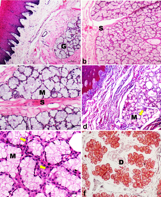
Histological Characteristics
Microscopic examination reveals distinct architectural patterns:
Glandular Structure
Porcine minor salivary glands exhibit compound tubuloalveolar organization. The secretory portions consist of:
- Simple cuboidal to pseudostratified epithelium
- Myoepithelial cells interspersed between secretory cells
- Well-developed basement membrane surrounding each acinus
- Interstitial connective tissue supporting the glandular architecture
Cellular Composition
The epithelial lining demonstrates:
- Basal cells – stem cell population for gland renewal
- Superficial cells – responsible for secretion production
- Dark cells – contain higher enzymatic activity
- Bright cells – primarily involved in mucin synthesis
Histochemical Analysis Findings
Special staining techniques reveal critical biochemical properties:
Secretory Product Identification
- Mucins – Periodic Acid-Schiff (PAS) positive, diastase-resistant
- Neutral carbohydrates – PAS positive, sensitive to diastase digestion
\li>Sulfated glycosaminoglycans – Alcian blue positive at pH 2.5
\li>Proteins – Cytochrome P450 detected in endoplasmic reticulum
Enzymatic Activity Patterns
- Amylase – Present in superficial acinar cells
- Lactate dehydrogenase – Widely distributed throughout parenchyma
- Glucose-6-phosphatase – Detected in basolateral cell membranes
\li>Alkaline phosphatase – Localized to basolateral surfaces
Comparative Analysis With Other Species
When compared to human and laboratory animal models:
Structural Similarities
- Compound tubuloalveolar arrangement conserved across species
- Similar epithelial cell stratification patterns
- Conserved myoepithelial cell distribution
\li>Key difference – Porcine glands tend to be larger with more abundant connective tissue stroma
Biochemical Variations
- Higher mucin production in porcine vs. human counterparts
- Differences in carbohydrate composition profiles
- Variations in enzyme distribution patterns
\li
Clinical relevance – These differences affect enzymatic assay interpretations in veterinary medicine
Veterinary and Research Applications
The porcine model offers distinct advantages:
- Anatomical similarity – Close resemblance to human oral cavity
- Physiological parallels – Comparable salivary flow rates and composition
- Research utility – Valuable for translational oral biology studies
- Pathological insights – Useful for comparative oral disease research
Conclusion
This comparative histological and histochemical analysis demonstrates that porcine minor salivary glands possess unique structural and biochemical characteristics while maintaining significant homology with other mammalian species. The compound tubuloalveolar architecture, combined with distinct enzymatic and glycoprotein profiles, establishes pigs as valuable models for oral gland research. These findings contribute to our understanding of oral physiology and provide foundational knowledge for veterinary dental practice and comparative medicine applications.
The integration of histological and histochemical approaches reveals the functional complexity underlying these seemingly simple exocrine structures, highlighting the importance of systematic anatomical investigation in veterinary science.
Comments are closed, but trackbacks and pingbacks are open.