Rapid Electrochemical Hemoglobin Detection in Mouse Feces Using a DNA Aptamer for Colitis Severity Assessment
Introduction
Colitis research often relies on invasive endoscopy or time‑consuming biochemical assays to gauge disease severity. A new rapid, non‑invasive method—electrochemical hemoglobin detection in mouse feces using a DNA aptamer—offers a convenient alternative. This technique provides real‑time insight into intestinal bleeding, a key indicator of colitis progression.
How the Aptamer‑Based Electrochemical Sensor Works
Key Components
- DNA aptamer: A short, single‑stranded oligonucleotide selected for high affinity to hemoglobin.
- Screen‑printed electrode (SPE): Low‑cost, disposable platform for signal transduction.
- Redox mediator: Facilitates electron transfer, amplifying the hemoglobin signal.
Detection Principle
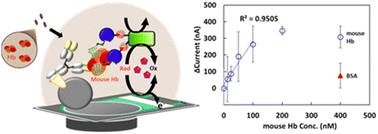
When fecal samples are applied to the SPE, hemoglobin binds to the immobilized aptamer. This complex modifies the electrode’s surface conductivity, producing a measurable current change proportional to hemoglobin concentration.
Step‑by‑Step Protocol for Mouse Feces
- Sample collection: Collect fresh fecal pellets, weigh 10–20 mg, and place in a microcentrifuge tube.
- Extraction: Add 200 µL phosphate‑buffered saline, vortex 30 seconds, and centrifuge 5 min at 10,000 g.
- Sensor preparation: Drop‑cast 5 µL of aptamer solution (1 µM) onto the working electrode and dry at room temperature for 10 minutes.
- Measurement: Pipette 10 µL of supernatant onto the electrode, insert into the handheld potentiostat, and record the square‑wave voltammetry signal.
- Data analysis: Compare the peak current to a calibrated standard curve (0–200 ng/µL hemoglobin) to quantify bleeding.
Why This Method Outperforms Traditional Approaches
- Speed: Complete analysis in under 5 minutes per sample.
- Non‑invasive: No need for blood draws or tissue biopsies.
- Cost‑effective: Reagents and disposable electrodes cost <$1 per test.
- Sensitivity: Detects hemoglobin as low as 5 ng/µL, sufficient for early‑stage colitis detection.
Application in a Mouse Model of Colitis
Researchers induced colitis in C57BL/6 mice with 3% dextran sodium sulfate (DSS) for 7 days. Fecal hemoglobin levels were measured daily using the aptamer sensor. Results showed a clear correlation:
- Day 0 (baseline): < 5 ng/µL.
- Day 3: 20–35 ng/µL, indicating mild mucosal damage.
- Day 7: >80 ng/µL, reflecting severe ulceration.
These values matched histopathology scores, confirming that fecal hemoglobin is a reliable surrogate marker for inflammation severity.
Integrating the Sensor into Your Research Workflow
Data Management Tips
- Log each mouse ID, weight, and DSS concentration alongside hemoglobin readings.
- Use simple spreadsheet formulas to calculate area‑under‑curve (AUC) for longitudinal studies.
Potential Modifications
- Combine with a multiplexed aptamer panel (e.g., calprotectin) for broader inflammatory profiling.
- Adapt the sensor for on‑site monitoring in animal facilities.
Conclusion
The DNA‑aptamer electrochemical sensor delivers rapid, inexpensive, and accurate hemoglobin detection in mouse feces, empowering researchers to evaluate colitis severity without invasive procedures. By adopting this method, labs can increase throughput, reduce animal stress, and obtain real‑time data that align closely with traditional histological assessments.
Comments are closed, but trackbacks and pingbacks are open.